Tunica vaginalis
Lua error in package.lua at line 80: module 'strict' not found.
| Tunica vaginalis | |
|---|---|
| File:Tunica vaginalis.jpg
Diagram of a cross-section of a testicle. 1. Cavity of tunica vaginalis, 2. Visceral lamina, 3. Parietal lamina.
|
|

The right testis, exposed by laying open the tunica vaginalis. (Tunica vaginalis is labeled at upper right.)
|
|
| Details | |
| Latin | tunica vaginalis testis |
| Identifiers | |
| Dorlands /Elsevier |
t_22/12832403 |
| TA | Lua error in Module:Wikidata at line 744: attempt to index field 'wikibase' (a nil value). |
| TH | {{#property:P1694}} |
| TE | {{#property:P1693}} |
| FMA | {{#property:P1402}} |
| Anatomical terminology
[[[d:Lua error in Module:Wikidata at line 863: attempt to index field 'wikibase' (a nil value).|edit on Wikidata]]]
|
|
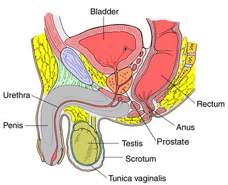
The tunica vaginalis is the serous covering of the testis. It is a pouch of serous membrane, derived from the processus vaginalis of the peritoneum, which in the fetus preceded the descent of the testis from the abdomen into the scrotum.[citation needed]
After its descent, that portion of the pouch which extends from the abdominal inguinal ring to near the upper part of the gland becomes obliterated; the lower portion remains as a shut sac, which invests the surface of the testis, and is reflected on to the internal surface of the scrotum; hence it may be described as consisting of a visceral and a parietal lamina.[citation needed]
Contents
Visceral lamina
The visceral lamina (lamina visceralis) covers the greater part of the testis and epididymis, connecting the latter to the testis by means of a distinct fold. From the posterior border of the gland it is reflected on to the internal surface of the scrotum.[citation needed]
Parietal lamina
The parietal lamina (lamina parietalis) is far more extensive than the visceral, extending upward for some distance in front and on the medial side of the cord, and reaching below the testis. The inner surface of the tunica vaginalis is smooth, and covered by a layer of simple squamous mesothelial cells. The interval between the visceral and parietal laminæ constitutes the cavity of the tunica vaginalis.[citation needed]
Diseases
- Mesothelioma
- Hydrocele[1]
- Cartilaginous Bodies[2]
- Hematocele[3]
Additional images
-

Schematic drawing of a cross-section through the vaginal process.

References
This article incorporates text in the public domain from the 20th edition of Gray's Anatomy (1918)Lua error in package.lua at line 80: module 'strict' not found.
- ↑ Hydrocele in Emergency Medicine at eMedicine
- ↑ Lua error in package.lua at line 80: module 'strict' not found.
- ↑ Lua error in package.lua at line 80: module 'strict' not found.
External links
- Diagram at aspiruslibrary.org
- Anatomy photo:36:st-1502 at the SUNY Downstate Medical Center - "Inguinal Region, Scrotum and Testes: Tunic"
- Swiss embryology (from UL, UB, and UF) ugenital/diffmorpho04
- inguinalregion at The Anatomy Lesson by Wesley Norman (Georgetown University) (testes)
- Articles to be merged from November 2013
- Pages with broken file links
- Medicine infobox template using GraySubject or GrayPage
- Medicine infobox template using Dorlands parameter
- Articles with unsourced statements from May 2015
- Wikipedia articles incorporating text from the 20th edition of Gray's Anatomy (1918)
- Articles that show a Medicine navs template
- Mammal male reproductive system